

Advanced Vibratome Technology for Life Science and Biomedical Research
The Campden 9000SMZ Vibrating Microtome is our most advanced system, engineered for precise sectioning of live and fixed brain tissue. Trusted by neuroscience, cardiac, lung, liver, organoid, and plant research labs worldwide, it delivers exceptional control, slice quality, and tissue viability—making it the ideal choice for electrophysiology, imaging, pathology, toxicology, and organ research.
A New Standard in Precision Tissue Slicing
Whether you’re preparing slices of brain, kidney, lung, liver, retina, cardiac, pancreatic, brain organoids, tumour, or plant tissue, the 9000SMZ provides the precision and reproducibility that today’s life sciences demand.
Superior Tissue Integrity Across Applications
- Ultra-low blade Z-axis deflection minimizes mechanical damage.
- Maintains structural and cellular viability in a wide range of soft tissues.
- Ideal for acute slicing, live tissue studies, ex vivo analysis, and long-term cultures.
Image: 40x IR-DIC Entorhinal Cortex Slice from 9 month-old Rat. Slicing Parameters: Blade: Ceramic; Blade Frequency: 50Hz; Blade Amplitude: 1mm; Advance Speed: 0.05 mm/s.

One Vibratome, Endless Applications

Key Features


Designed for Scientists, Backed by Innovation
Campden Instruments has collaborated with leading researchers across various research disciplines to build a vibratome that meets real-world laboratory challenges. Find confidence and precision with the 9000SMZ.
Customer Spotlights
Dr. Gareth Morris of University of Manchester and University College London talks about his research and how the use of Brain Slices provides an invaluable, in vitro research tool.
Dr. Kate Connor from Trinity College Dublin talks about her research into the development of clinically relevant cancer models and the use of brain slices for model validation.
Example Data
Oscillations in slices from 3 week-old mice
Neuronal oscillations in the theta frequency range (4 – 8 Hz) induced by the acetylcholine muscarinic receptor agonist carbachol (50 μM) in hippocampal area CA3 from 3 week-old C57BL/6J mice. Slices were either incubated in control aCSF or in aCSF supplemented with sonicated recombinant human tau preformed fibrils (133 nM; rPeptide; CF-1001-1).
Left panels: raw electrophysiological traces showing induction of oscillations after carbachol, and 25 s of activity (red boxed region; lower traces).
Right panels: spectrograms of theta activity induced by carbachol in both conditions.
Source: Adwoa Boaten, Ben Hopkins, Tegan Lawrence, Beth Rees, Amol Bhandare, Stuart Greenhill, Gavin Woodhall, Mark Wall, Bruno Frenguelli School of Life Sciences, University of Warwick, Coventry, UK CV4 7AL. Institute of Health and Neurodevelopment, Aston University, Birmingham, UK B4 7ET

Oscillations in slices from 9 month-old rats
Improved microtome design allows neuronal preservation and physiological oscillations in older rats (9 months). Here we show DIC images from entorhinal cortex and area CA1.
(A) Raw electrophysiological data at increasing magnification demonstrating characteristic spontaneous sawtooth gamma oscillations in LFP recordings (450 μm slice).
(B) Panel shows example FFT from LFP recording in hippocampus with no added drugs, with high power in gamma range and excellent coherence.
(C) Whole-cell patch-clamp recording from hippocampal cell with regular EPSCs and stable baseline (350 μm slice).

Immunohistochemistry

Maximum intensity projections of confocal images showing choline acetyltransferase (ChAT)+ neurons (left, yellow) and glial fibrillary acidic protein (GFAP)+ astroglia (right, red) in40 μm thick mouse brainstem sections. Scale bars = 20 μm

Left: Iba1 staining of LPS-stimulated microglia in the CA1 region of a 200 μm hippocampal slice. Right: highlighted region of interest. Scale bars = 50 μm
Source: Adwoa Boaten, Ben Hopkins, Tegan Lawrence, Beth Rees, Amol Bhandare, Stuart Greenhill, Gavin Woodhall, Mark Wall, Bruno Frenguelli School of Life Sciences, University of Warwick, Coventry, UK CV4 7AL. Institute of Health and Neurodevelopment, Aston University, Birmingham, UK B4 7ET
Specifications:
Product Resources
Product Manuals
Research Papers and Bibliographies
Related Products
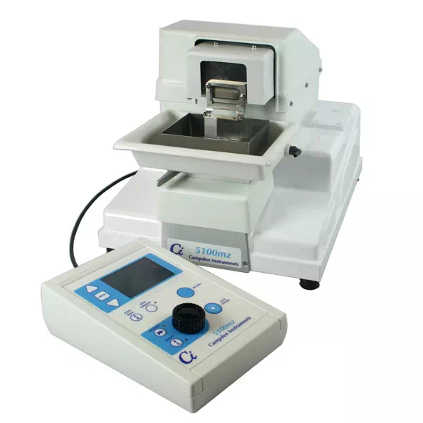
5100mz Vibratome
The 5100mz is a very competitively priced, high precision, vibrating microtome (vibrotome for short) which shares many features with the top of the range 7000smz series, such as the vibrating mechanism, the inner and outer tissue baths and the easy to use control system.
View Details5100mz-Plus Vibratome
The 5100-Plus is perfect for those who need to keep slices viable for longer e.g. for electrophysiological field recordings. The user can calibrate the Z-axis deflection of the blade to 2 µm with the adjustable blade holder and “Opti-cal” Calibration device.
View Details